Human M-CSF ELISA Kit
Size: 96T
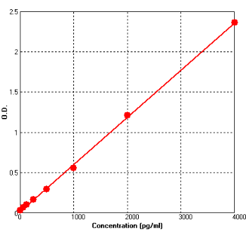
Range: 62.5 pg/ml-4000 pg/ml
Sensitivity < 10pg/ml
Application: For quantitative detection of M-CSF in tissue lysates or cell culture supernates, human serum and plasma should be validated by end users.
--------------------------------------------------------------------------------------------------------------
Price: ---
Catalog No.: BEK1146
Size: 96T
Range: 62.5 pg/ml-4000 pg/ml
Sensitivity < 10pg/ml
Storage and Expiration: Store at 2-8℃ for 6 months, or at -20℃ for 12 months.
Application: For quantitative detection of M-CSF in tissue lysates or cell culture supernates, human serum and plasma should be validated by end users.
Introduction
Macrophage colony-stimulating factor, or M-CSF, is a secreted cytokine which influences hematopoietic stem cells to differentiate into macrophages or other related cell types. Ladner et al. (1987) showed that there are 2 forms of M-CSF, with 224 and 522 amino acids, resulting from alternative splicing. It is a hematopoietic growth factor that is involved in the proliferation, differentiation, and survival of monocytes, macrophages, and bone marrow progenitor cells. It released by osteoblasts (as a result of endocrine stimulation by parathyroid hormone) exerts paracrine effects on osteoclasts. M-CSF binds to receptors on osteoclasts inducing differentiation, and ultimately leading to increased plasma calcium levels—through the resorption (breakdown) of bone. More recently, it was discovered that CSF-1 and its receptor CSF1R are implicated in the mammary gland during normal development and neoplastic growth.
Principle of the Assay
This kit was based on sandwich enzyme-linked immune-sorbent assay technology. Anti-M-CSF polyclonal antibody was pre-coated onto 96-well plates. And the biotin conjugated anti-M-CSF polyclonal antibody was used as detection antibodies. The standards, test samples and biotin conjugated detection antibody were added to the wells subsequently, and wash with wash buffer. Avidin-Biotin-Peroxidase Complex was added and unbound conjugates were washed away with wash buffer. TMB substrates were used to visualize HRP enzymatic reaction. TMB was catalyzed by HRP to produce a blue color product that changed into yellow after adding acidic stop solution. The density of yellow is proportional to the M-CSF amount of sample captured in plate. Read the O.D. absorbance at 450nm in a microplate reader, and then the concentration of M-CSF can be calculated.
Kit components
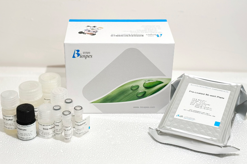
- One 96-well plate pre-coated with anti-Human M-CSF antibody
- Lyophilized Human M-CSF standards: 2 tubes (10ng / tube)
- Sample / Standard diluent buffer: 30ml
- Biotin conjugated anti-Human M-CSF antibody (Concentrated): 130μl. Dilution: 1:100
- Antibody diluent buffer: 12ml
- Avidin-Biotin-Peroxidase Complex (ABC) (Concentrated): 130μl. Dilution: 1:100
- ABC diluent buffer: 12ml
- TMB substrate: 10ml
- Stop solution: 10ml
- Wash buffer (25X): 30ml
Note: Reconstitute standards and test samples with Kit Component 3.
Material Required But Not Provided
- 37℃ incubator
- Microplate reader (wavelength: 450nm)
- Precise pipette and disposable pipette tips
- Automated plate washer
- ELISA shaker
- 1.5ml of Eppendorf tubes
- Plate cover
- Absorbent filter papers
- Plastic or glass container with volume of above 1L
Details
Product Center
